The Imaging Center at ATLATL is designed to help teams visualize biology, validate mechanisms, and generate decision-ready data quickly.
By integrating advanced microscopy, high-content imaging, and in vivo imaging systems, the platform enables researchers to study biological processes across molecular, cellular, and whole-animal levels. From intracellular dynamics to tumor progression, the platform provides high-resolution, real-time insights that accelerate discovery and translational research.
Located within ATLATL’s bio-hubs, scientists work side-by-side with imaging experts, enabling rapid experiment setup, real-time troubleshooting, and faster iteration. By combining imaging with downstream analysis, teams can move efficiently from observation to actionable conclusions.
Core Capabilities:
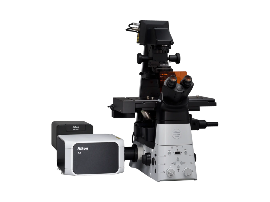
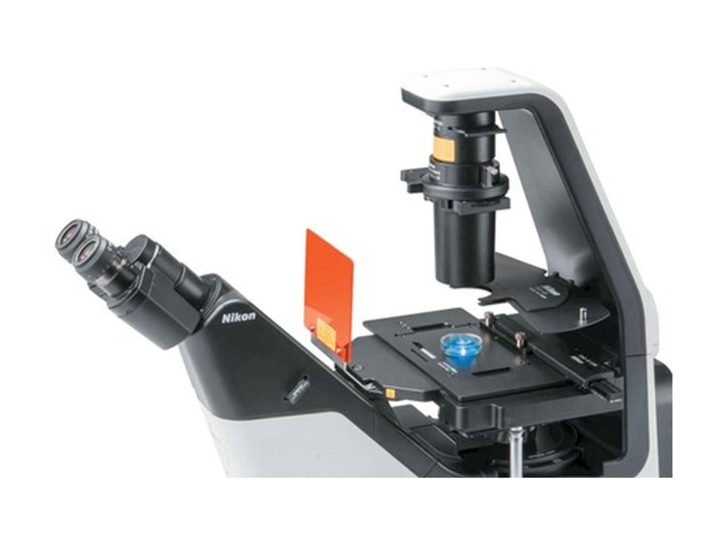
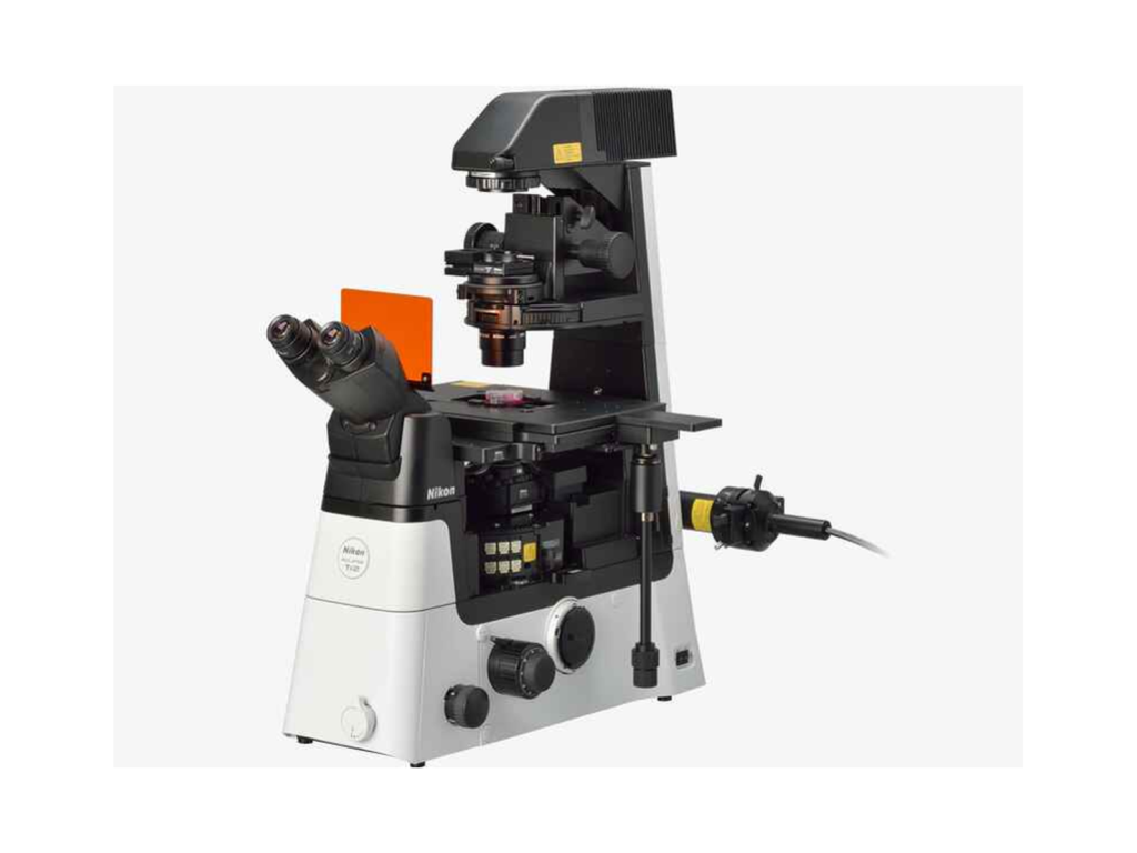
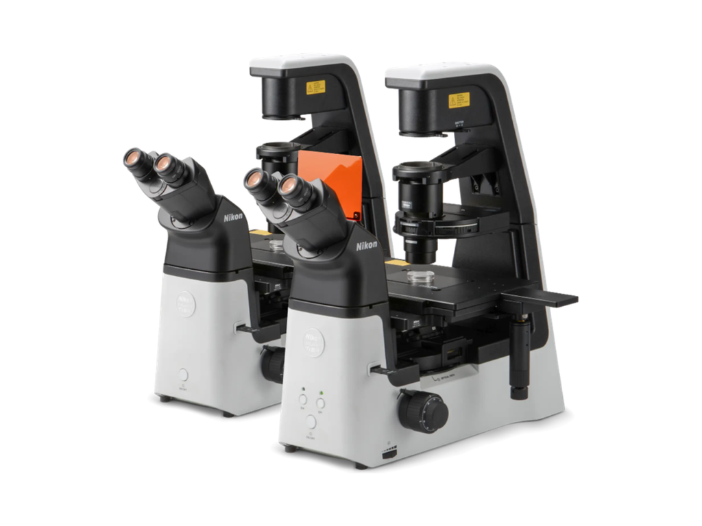
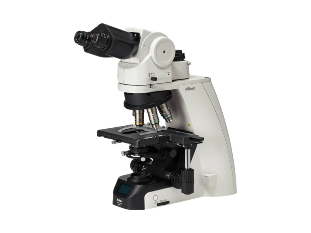
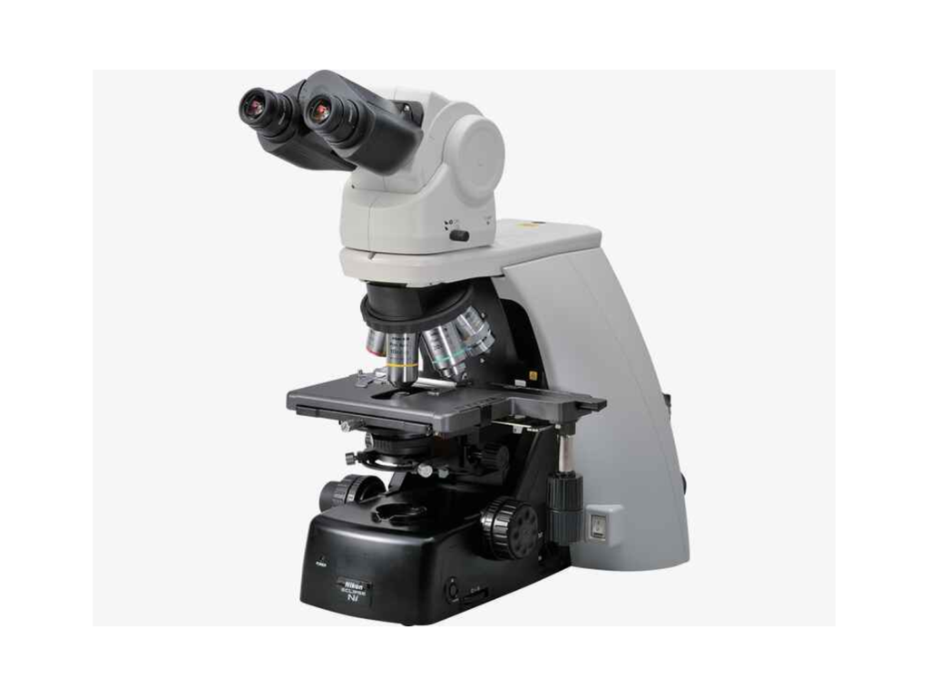
Advanced Microscopy & Live-Cell Imaging
High-resolution, multicolor imaging of cells in 2D and 3D systems, including neurons, cancer cells, and complex models.
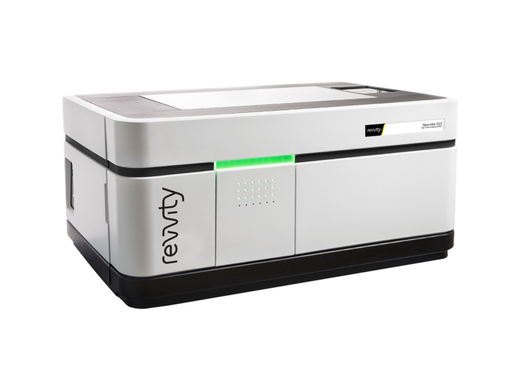
High-Content & Phenotypic Imaging
Automated imaging workflows for quantitative, high-throughput analysis, supporting screening and functional studies.
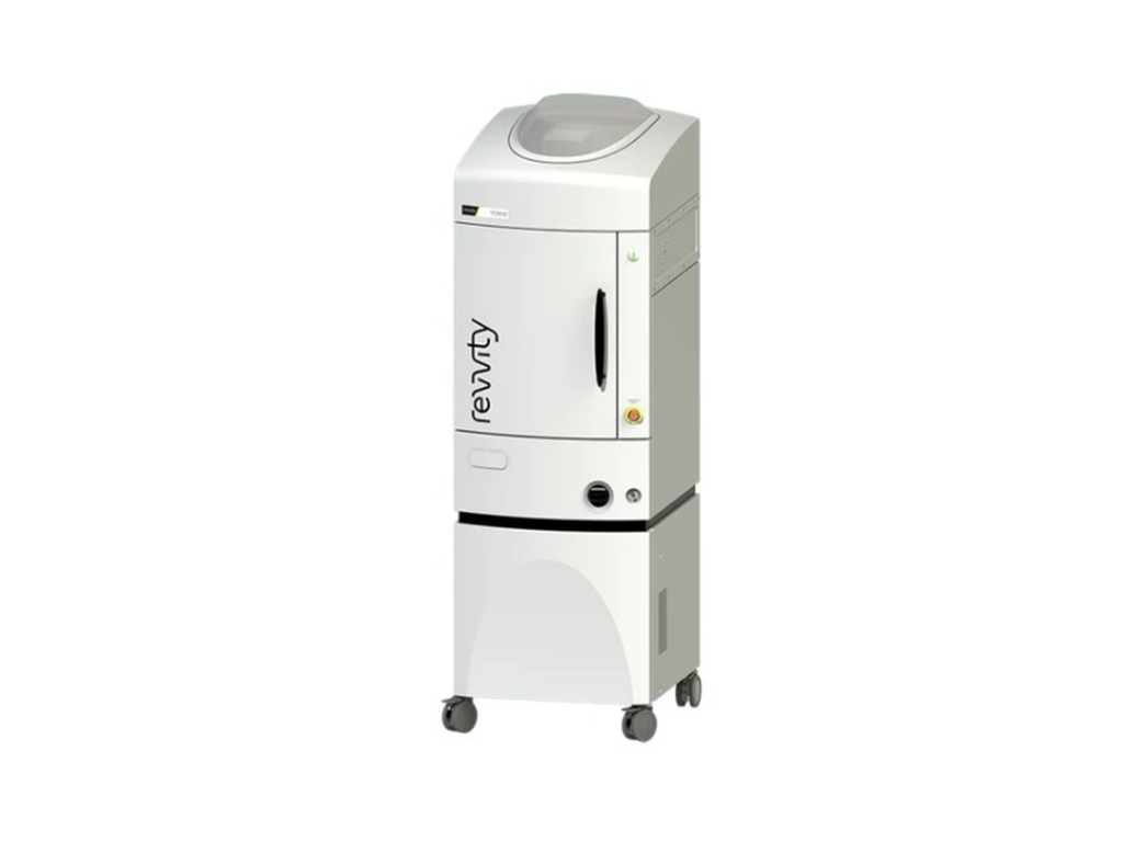
In Vivo Imaging
Real-time, non-invasive imaging using IVIS systems, enabling longitudinal studies in animal models.
Single-Molecule & Mechanistic Analysis
Tracking molecular behavior and intracellular dynamics to understand mechanism of action at high resolution.
Laser Microsurgery & Cellular Manipulation
Precision tools for cell ablation and manipulation to study function and biological responses.
Equipment:
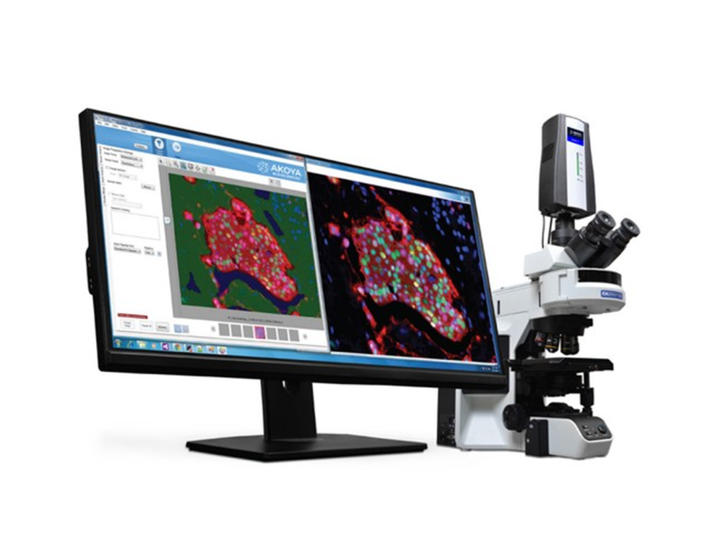
AkoyaBio Mantra 2™ Quantitative Pathology Imaging and Analysis
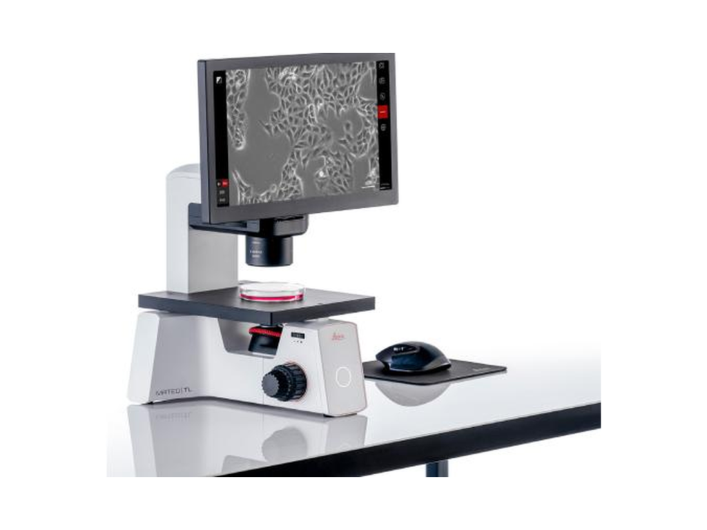
Leica Mateo TL digital transmitted light inverted microscope




 English
  日本語
日本語
 简体中文
简体中文
 Français
 Deutsch